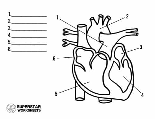
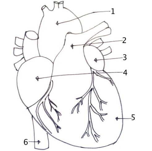
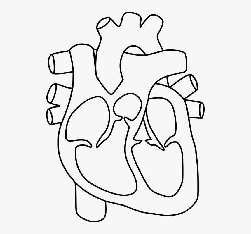
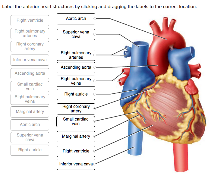
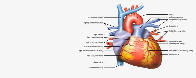
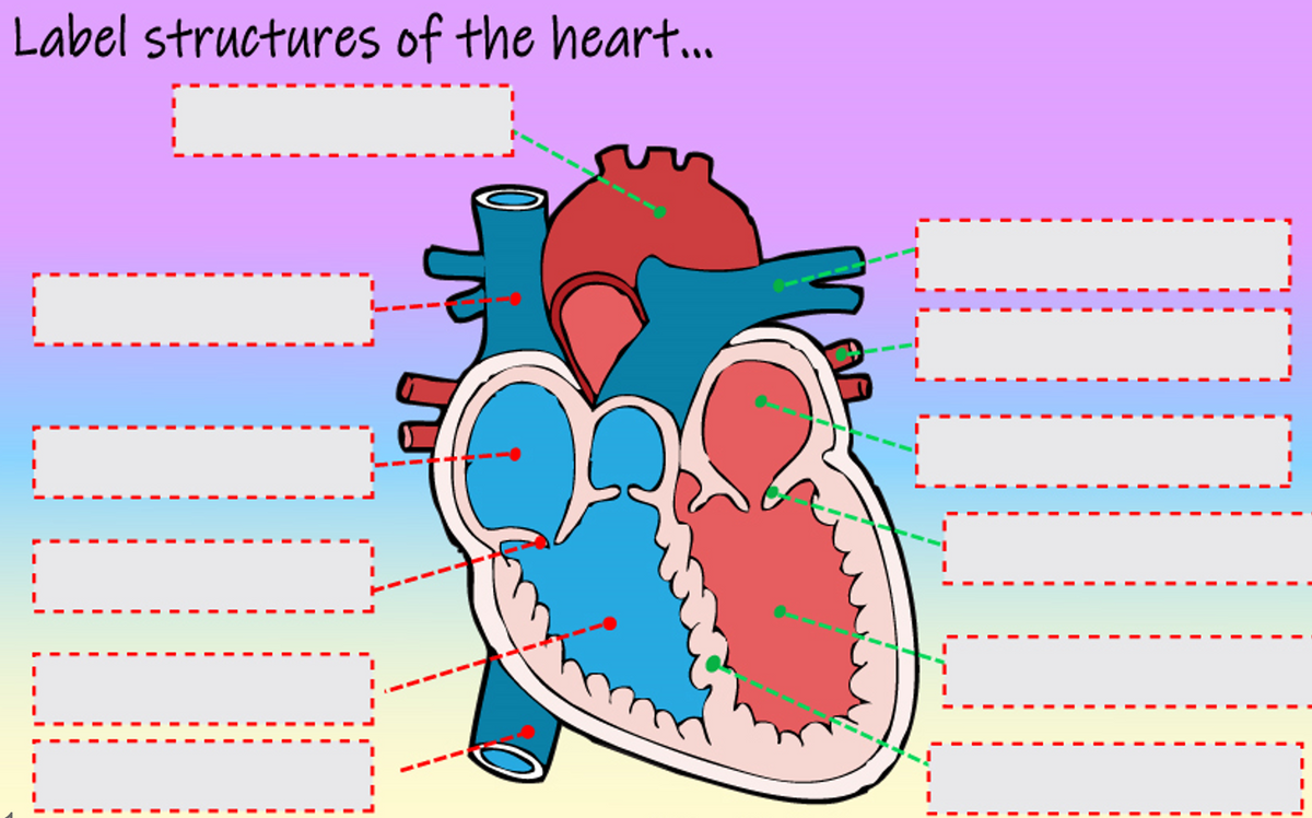
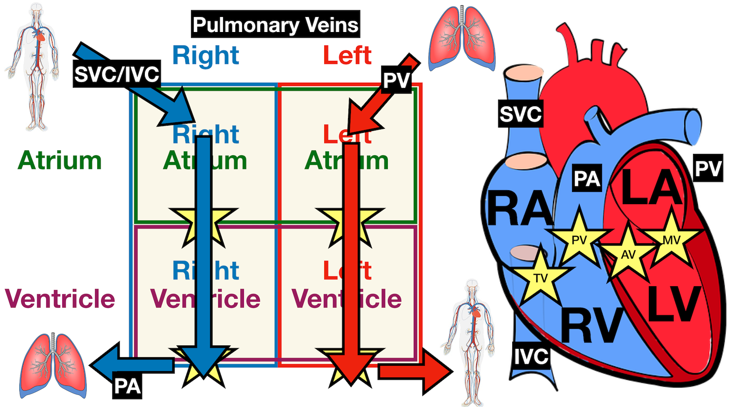
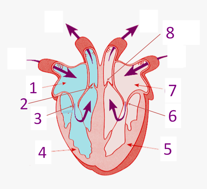
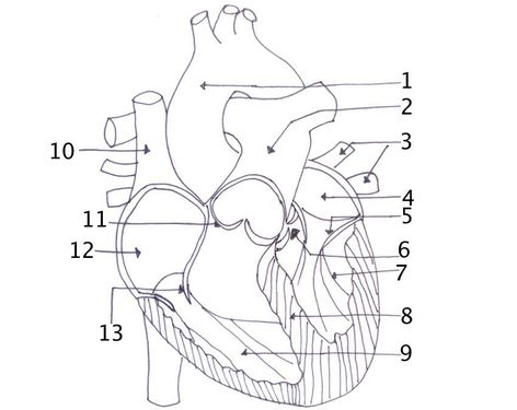
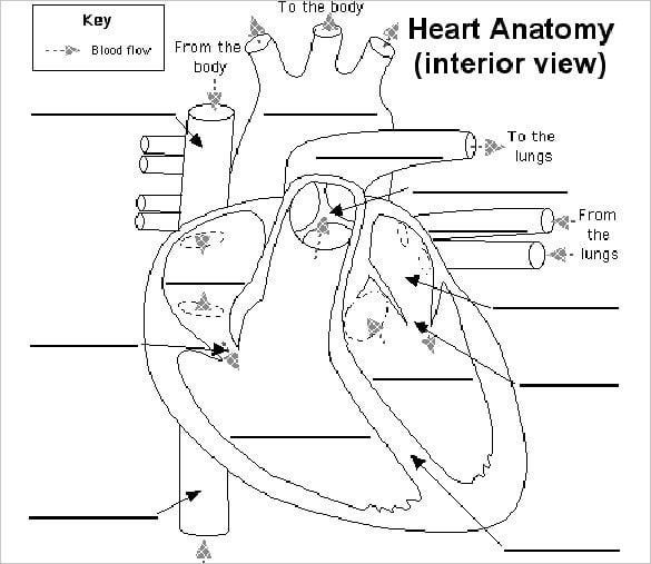
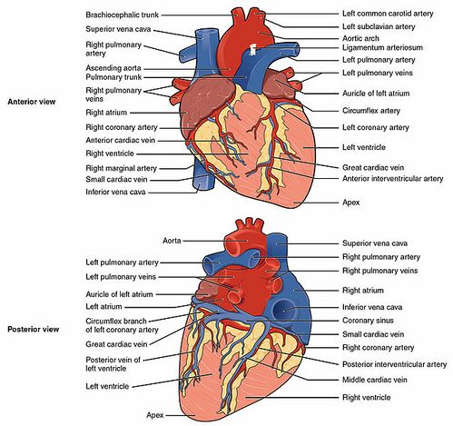
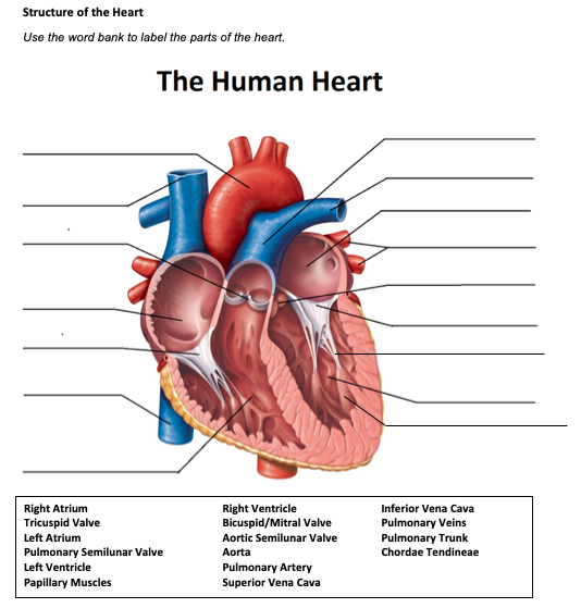
41 structure of the heart without labels
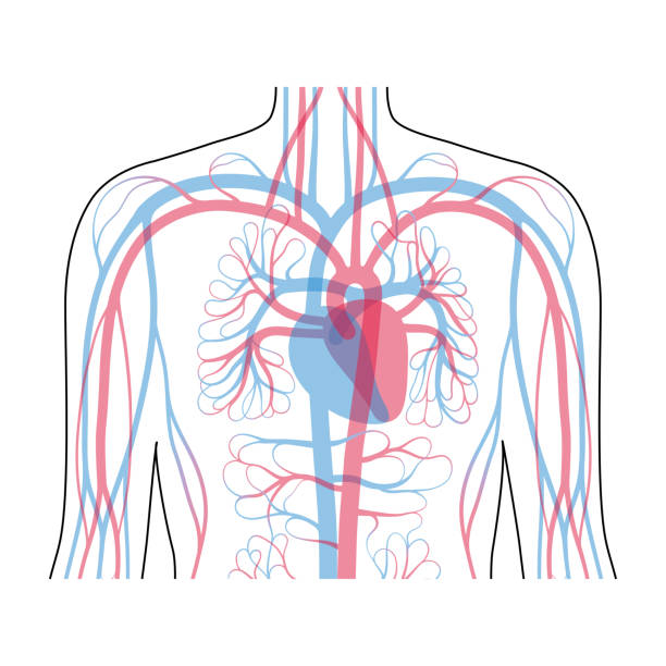
Circulatory system - Wikipedia The circulatory system includes the heart, blood vessels, and blood. The cardiovascular system in all vertebrates, consists of the heart and blood vessels. The circulatory system is further divided into two major circuits - a pulmonary circulation, and a systemic circulation. The pulmonary circulation is a circuit loop from the right heart taking deoxygenated blood to the lungs where it is ... Fellow of the American Heart Association (FAHA) For those who qualify, election as a Fellow of the American Heart Association recognizes your scientific and professional accomplishments, volunteer leadership and service. By earning the right to include the initials FAHA among your credentials, you let colleagues and patients know that you have been welcomed into one of the world’s most eminent organizations of …
› books › NBK526065Losartan - StatPearls - NCBI Bookshelf Jul 18, 2022 · Losartan is FDA approved for the treatment of several medical conditions, which include the following: hypertension, diabetic nephropathy. ARBs are known to be renoprotective in type 2 diabetes mellitus. In hypertension with left ventricular hypertrophy, losartan inhibits angiotensin II-induced cardiac remodeling. It reduces the risk of stroke in these patients. This activity covers losartan ...

Structure of the heart without labels
Capillaries: Anatomy, Function, and Significance - Verywell Health Capillaries are found throughout the body and may be thought of as the central portion of circulation. Blood leaves the heart through the aorta and the pulmonary arteries, traveling to the rest of the body and to the lungs, respectively. These large arteries become smaller arterioles and eventually narrow to form the capillary bed. Structure And Function Of The Heart (Post Test) - ProProfs Quiz Right atrium → right ventricle → lungs → left atrium → left ventricle. C. Left atrium → left ventricle → lungs → right ventricle → right atrium. D. Left atrium → left ventricle → right ventricle → right atrium → lungs. 5. A structure in the heart that temporarily closes, ensuring that blood moves in only one direction. A. human body | Organs, Systems, Structure, Diagram, & Facts Thus, the heart is an organ composed of all four tissues, whose function is to pump blood throughout the body. Of course, the heart does not function in isolation; it is part of a system composed of blood and blood vessels as well. The highest level of body organization, then, is that of the organ system.
Structure of the heart without labels. Metal Hammer | Louder Vor 2 Tagen · Death, floods and therapy: how Parkway Drive found their way out of the darkness Parkway Drive have spent the last few years living their own Some Kind Of Monster-style nightmare. Now they’re looking for the light at the end of the tunnel Layers of the heart: Epicardium, myocardium, endocardium - Kenhub The epicardium is the outermost layer of the heart. It is actually the visceral layer of the serous pericardium, which adheres to the myocardium of the heart. Histologically, it is made of mesothelial cells, the same as the parietal pericardium. Heart: illustrated anatomy - e-Anatomy - IMAIOS Morphology - pericardium Myocardium - fibrous structures Left cavities Right cavities Valves : aortic, mitral tricuspid and pulmonary valves Left coronary artery Right coronary artery Veins Conduction system Mediastium vessels such as aorta, pulmonary arteries and veins, superior vena cava. Pathology Outlines - Histology Newly oxygenated blood returns to the left atrium via 4 pulmonic veins at a normal pressure of approximately 10 mmHg. Left atrial contraction moves blood through the mitral valve into the left ventricle. Pacemaker of the heart located near the sinotubular junction, where the superior vena cava meets the right atrium.
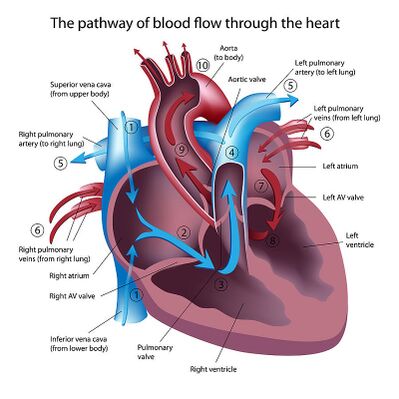
Parts of the Heart: How does blood flow through it? - Study.com Four hollowed-out parts of the heart are known as chambers. These chambers are situated such that there are two chambers on top and two on the bottom of the heart. Two of them are on the right side... furosemide: Edema Uses, Side Effects & Dosage - MedicineNet 10.08.2022 · Furosemide is a drug used to treat excessive fluid accumulation and swelling (edema) of the body caused by heart failure, cirrhosis, chronic kidney failure, and nephrotic syndrome. Common side effects of furosemide are low blood pressure, dehydration and electrolyte depletion (for example, sodium, potassium). Do not take if breastfeeding. Consult … The Ear: Anatomy, Function, and Treatment - Verywell Health The middle ear (also known as the tympanum or tympanic cavity) is a complicated network of tunnels, chambers, openings, and canals mostly inside openings within the temporal bone on each side of the skull. The 2 largest chambers are called the middle ear space and mastoid. Human heart: Anatomy, function & facts | Live Science The heart's outer wall consists of three layers. The outermost wall layer, or epicardium, forms the inner wall of the pericardium. The middle layer, or myocardium, contains the muscle that...
How the Heart Works - The Heart | NHLBI, NIH - National Institutes of ... The heart is an organ about the size of your fist that pumps blood through your body. It is made up of multiple layers of tissue. Your heart is at the center of your circulatory system. This system is a network of blood vessels, such as arteries, veins, and capillaries, that carries blood to and from all areas of your body. Losartan - StatPearls - NCBI Bookshelf 18.07.2022 · Losartan is FDA approved for the treatment of several medical conditions, which include the following: hypertension, diabetic nephropathy. ARBs are known to be renoprotective in type 2 diabetes mellitus. In hypertension with left ventricular hypertrophy, losartan inhibits angiotensin II-induced cardiac remodeling. It reduces the risk of stroke in these patients. This … Normal CT chest lung on axial images with labels | e-Anatomy - IMAIOS Arteries of the mediastinum Veins of the mediastinum Muscles and layers of the thoracic cavity. Bones of the thorax Nerves As the cursor is moved over an anatomical area of the lung parenchyma, the segment is highlighted and labeled: this tool was chosen to show segmental lung anatomy (international and Ikeda). Heart anatomy: Structure, valves, coronary vessels | Kenhub The heart is shaped as a quadrangular pyramid, and orientated as if the pyramid has fallen onto one of its sides so that its base faces the posterior thoracic wall, and its apex is pointed toward the anterior thoracic wall.
Analogy - Wikipedia Analogy (from Greek analogia, "proportion", from ana-"upon, according to" [also "against", "anew"] + logos "ratio" [also "word, speech, reckoning"]) is a cognitive process of transferring information or meaning from a particular subject (the analog, or source) to another (the target), or a linguistic expression corresponding to such a process. . In a narrower sense, analogy is an …
How the Heart Works: Diagram, Anatomy, Blood Flow - MedicineNet The heart is located under the rib cage -- 2/3 of it is to the left of your breastbone (sternum) -- and between your lungs and above the diaphragm. The heart is about the size of a closed fist, weighs about 10.5 ounces, and is somewhat cone-shaped. It is covered by a sack termed the pericardium or pericardial sack.
SGLT2 inhibitor - Wikipedia SGLT2 inhibitors, also called gliflozins or flozins, are a class of medications that modulate sodium-glucose transport proteins in the nephron (the functional units of the kidney), unlike SGLT1 inhibitors that perform a similar function in the intestinal mucosa.The foremost metabolic effect of this is to inhibit reabsorption of glucose in the kidney and therefore lower blood sugar.
15 Best Anatomy Apps 2022 (Android & iOS) - Freeappsforme They look very realistic. The app has 6 large sections such as the heart, brain, reproductive system, respiratory system, digestive system, and others. The app can show all anatomical systems together at once. The app has an animation of the movement of organs. You can remove the layers to look at the internal structure of the organs.
Structure/Function Claims | FDA Structure/function claims may describe the role of a nutrient or dietary ingredient intended to affect the normal structure or function of the human body, for example, "calcium builds strong bones ...
The Location, Size, and Shape of the Heart | GetBodySmart The Location, Size, and Shape of the Heart. The heart is located underneath the sternum in a thoracic compartment called the mediastinum, which occupies the space between the lungs. The sternum and mediastinum. It is approximately the size of a man's fist (230-350 grams) and is shaped like an inverted cone. About two thirds of the heart's ...
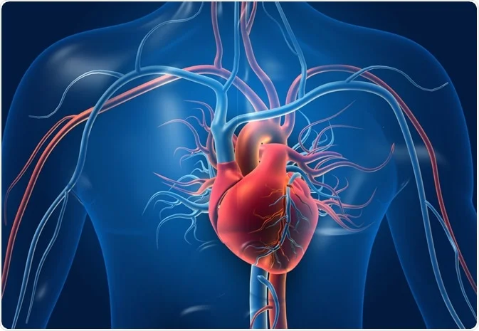
Structure and Function of the Heart - News-Medical.net Structure of the heart The heart wall is composed of three layers, including the outer epicardium (thin layer), middle myocardium (thick layer), and innermost endocardium (thin layer). The...
professional.heart.org › en › partnersFellow of the American Heart Association (FAHA) International applicants will be required to provide the same online application data as domestic applicants. Letter of Recommendation. If the candidate does not have access to a FAHA who knows their work, their letter of recommendation may be authored by the Chair or Academic Chair of their institution or by an international scientific leader.
Heart Diagram – 15+ Free Printable Word, Excel, EPS, PSD For every use a template has been designed with a motive of making it easy for the user to get the print of it without making a new one of his own. You may also visit venn diagram templates. Teachers and students use the heart diagram, in biological science, to study the structure and functions of a human being’s heart.
What Are the Four Main Functions of the Heart? - MedicineNet The heart is a muscular organ situated in the chest just behind and slightly toward the left of the breastbone. It roughly measures the size of a closed fist. The heart works all the time, pumping blood through the network of blood vessels called the arteries and veins. The heart and its blood vessels are known as the cardiovascular system.
Mnemonics for Heart Anatomy and Physiology (Video) - Mometrix Each half of the heart has an upper collecting chamber, the atrium, and a lower pumping chamber, the ventricle. You can remember their location because A comes before V. The atrium is above the ventricle. Heart Sounds Next, we'll take a look at the heart sounds heard with a stethoscope.
Structure of Cell: Definition, Types, Diagram, Functions - Embibe The nucleus is chemically composed of proteins, DNA, RNA and lipids. The shape of the nucleus may be spherical, oval or discoidal. The nucleus in plant cells is lens-shaped and peripheral in position due to a large central vacuole. It is spherical in animal cells and is located at the centre.
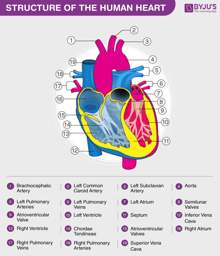
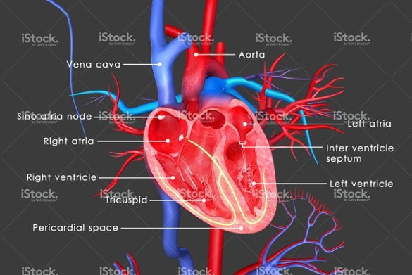
heart | Structure, Function, Diagram, Anatomy, & Facts The heart consists of several layers of a tough muscular wall, the myocardium. A thin layer of tissue, the pericardium, covers the outside, and another layer, the endocardium, lines the inside. The heart cavity is divided down the middle into a right and a left heart, which in turn are subdivided into two chambers.
Why the world of LGBTQ health doesn't fit under a single label The latest Gallup poll combined with census data puts the number of LGBTQ adults living in the U.S. at around 18 million, Streed said. So it might be obvious that broad labels would not apply to all. But Streed, who also is the research lead for the Center for Transgender Medicine and Surgery at Boston Medical Center, said that not only is each ...
Free Skeletal System Worksheets and Printables - Homeschool Giveaways The axial skeleton includes the 80 bones along the body's vertical axis such as the rib cage, spine, and skull. It provides protection and support for the spinal cord, brain, and internal organs such as the stomach, lungs, and heart. The appendicular skeleton is made up of the remaining 126 bones that attach to the axial skeleton.
Heart Labeling Quiz: How Much You Know About Heart Labeling? Here is a Heart labeling quiz for you. The human heart is a vital organ for every human. The more healthy your heart is, the longer the chances you have of surviving, so you better take care of it. Take the following quiz to know how much you know about your heart. Questions and Answers 1. What is #1? 2. What is #2? 3. What is #3? 4. What is #4?
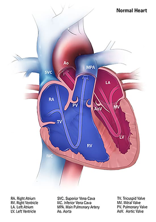
Heart - Wikipedia The heart has four chambers, two upper atria, the receiving chambers, and two lower ventricles, the discharging chambers.The atria open into the ventricles via the atrioventricular valves, present in the atrioventricular septum.This distinction is visible also on the surface of the heart as the coronary sulcus. There is an ear-shaped structure in the upper right atrium called the right atrial ...
› furosemide › articlefurosemide: Edema Uses, Side Effects & Dosage - MedicineNet Aug 10, 2022 · Furosemide is a drug used to treat excessive fluid accumulation and swelling (edema) of the body caused by heart failure, cirrhosis, chronic kidney failure, and nephrotic syndrome. Common side effects of furosemide are low blood pressure, dehydration and electrolyte depletion (for example, sodium, potassium). Do not take if breastfeeding. Consult your doctor if pregnant.
› design-templates › printHeart Diagram – 15+ Free Printable Word, Excel, EPS, PSD ... For every use a template has been designed with a motive of making it easy for the user to get the print of it without making a new one of his own. You may also visit venn diagram templates. Teachers and students use the heart diagram, in biological science, to study the structure and functions of a human being’s heart.
Pericardium - Structure & Function | GetBodySmart Tutorial: Surrounding the heart is a fibrous sac called the pericardium ( Gr., peri, around + kardia, heart ), which performs several functions. Fluids within the sac lubricate the outer wall of the heart so it can beat without causing friction. It also holds the heart in place, forms a barrier against infections, and helps keeps the heart from ...
Path of Blood Through the Heart | New Health Advisor Basics Parts of the Heart. Understanding the function of the heart is helpful to learn more about its anatomy. Here are the basic parts of the heart: 1. Right Atrium. The heart can be divided into right and left halves, as well as into the upper and lower chambers. There are two upper chambers called atria and two lower chambers called ventricles.
Diagram of Human Heart and Blood Circulation in It Four Chambers of the Heart and Blood Circulation The shape of the human heart is like an upside-down pear, weighing between 7-15 ounces, and is little larger than the size of the fist. It is located between the lungs, in the middle of the chest, behind and slightly to the left of the breast bone.
human body | Organs, Systems, Structure, Diagram, & Facts Thus, the heart is an organ composed of all four tissues, whose function is to pump blood throughout the body. Of course, the heart does not function in isolation; it is part of a system composed of blood and blood vessels as well. The highest level of body organization, then, is that of the organ system.
Structure And Function Of The Heart (Post Test) - ProProfs Quiz Right atrium → right ventricle → lungs → left atrium → left ventricle. C. Left atrium → left ventricle → lungs → right ventricle → right atrium. D. Left atrium → left ventricle → right ventricle → right atrium → lungs. 5. A structure in the heart that temporarily closes, ensuring that blood moves in only one direction. A.
Capillaries: Anatomy, Function, and Significance - Verywell Health Capillaries are found throughout the body and may be thought of as the central portion of circulation. Blood leaves the heart through the aorta and the pulmonary arteries, traveling to the rest of the body and to the lungs, respectively. These large arteries become smaller arterioles and eventually narrow to form the capillary bed.















Post a Comment for "41 structure of the heart without labels"