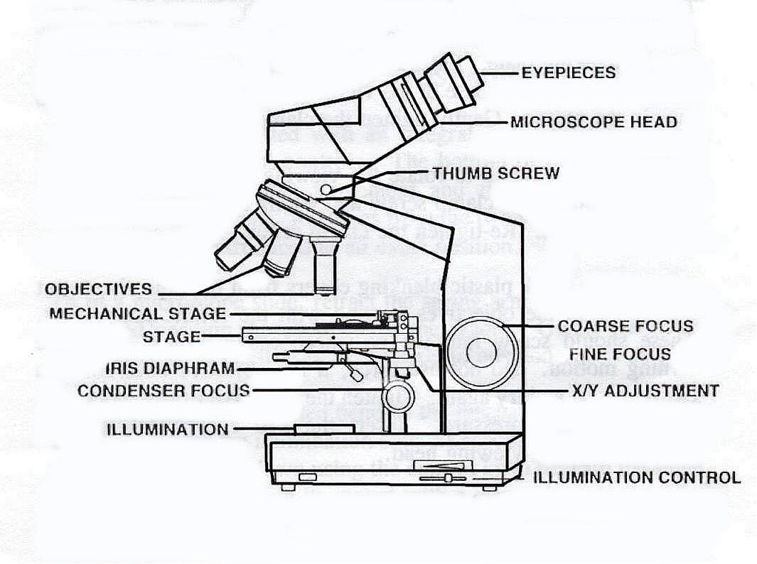
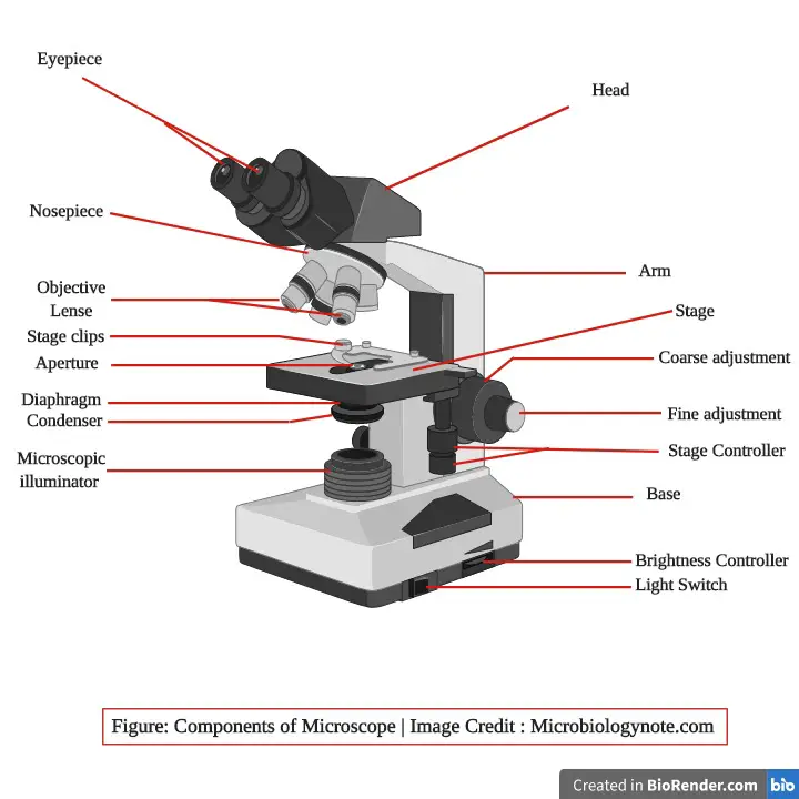
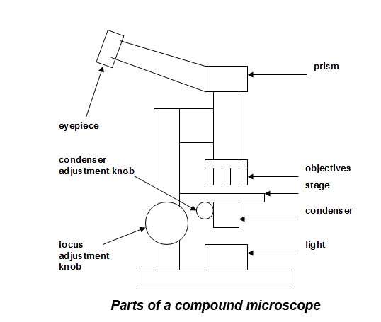
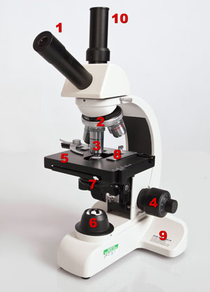
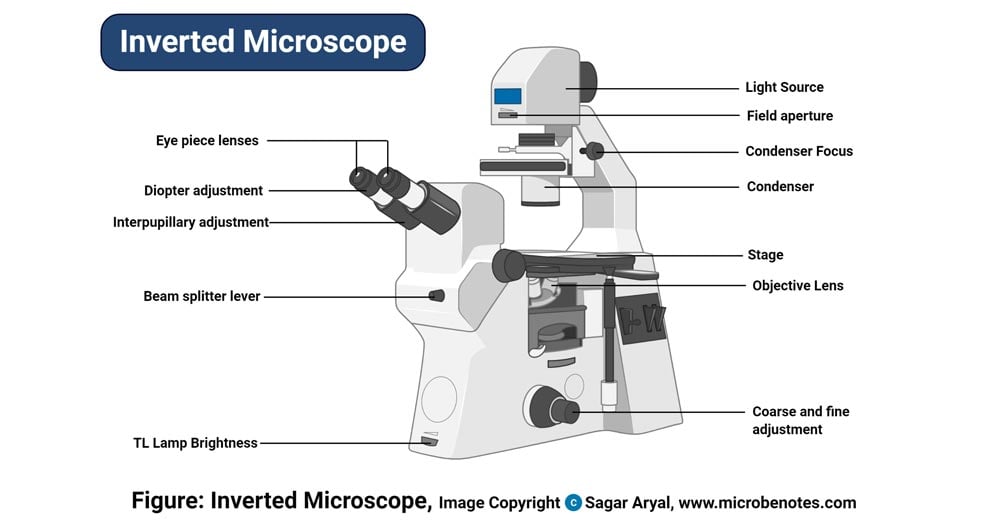
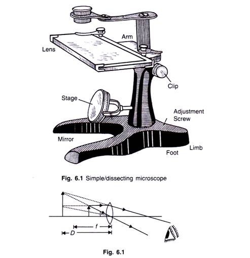
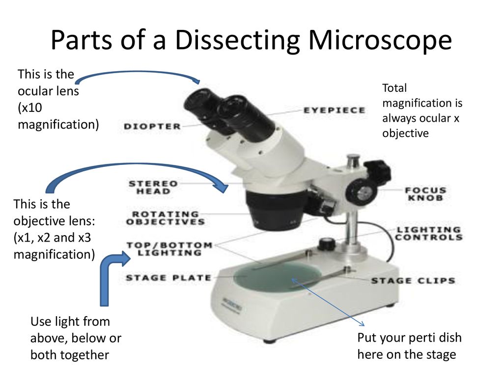
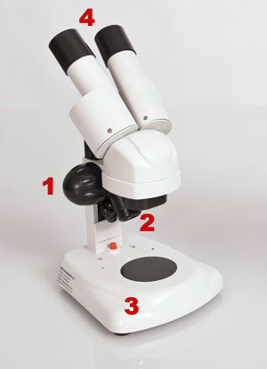
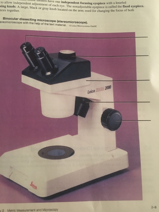
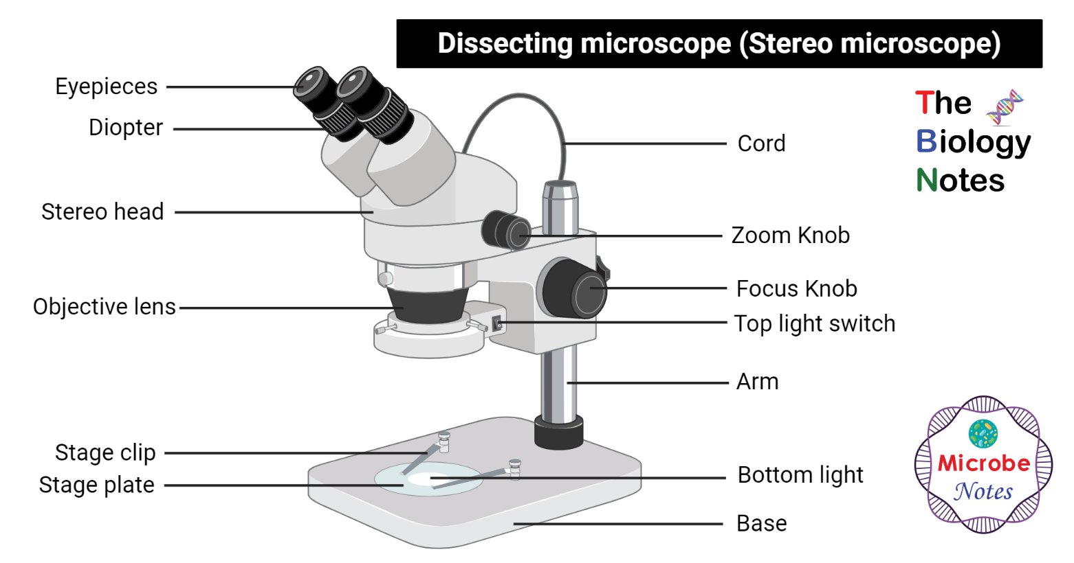
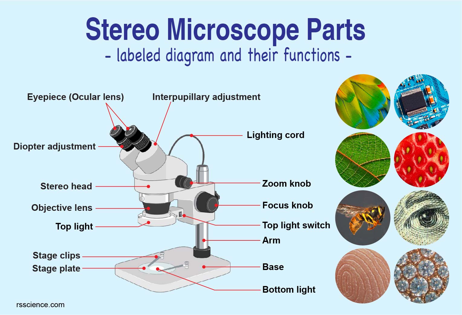
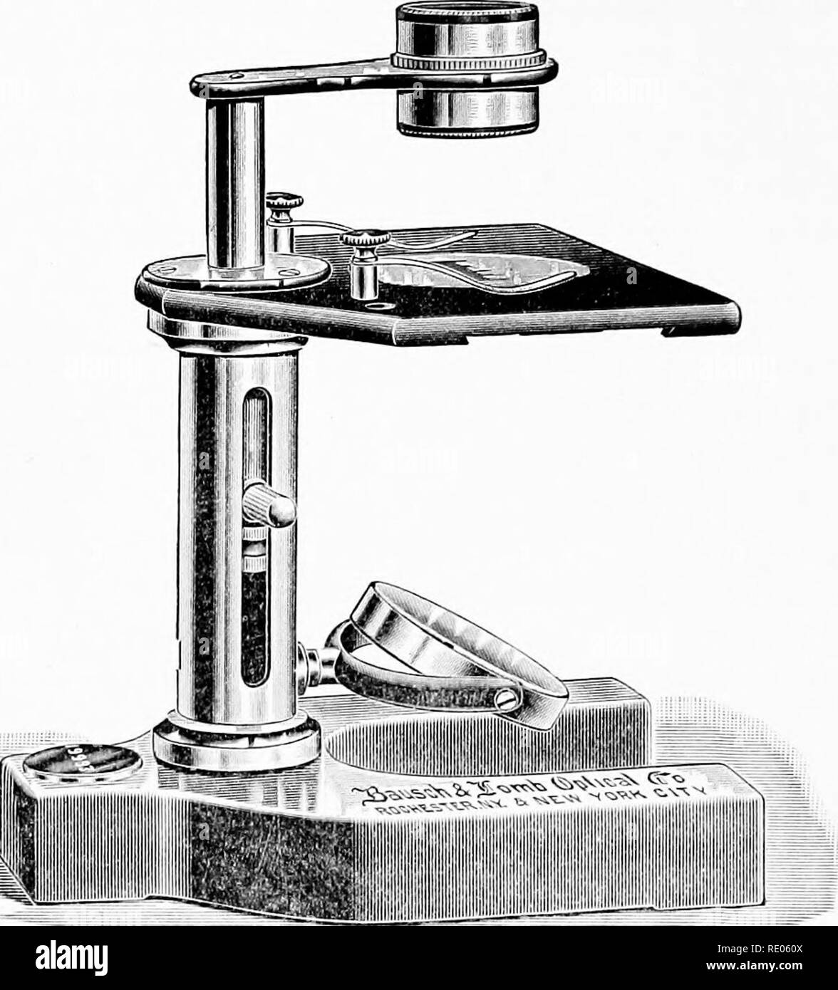
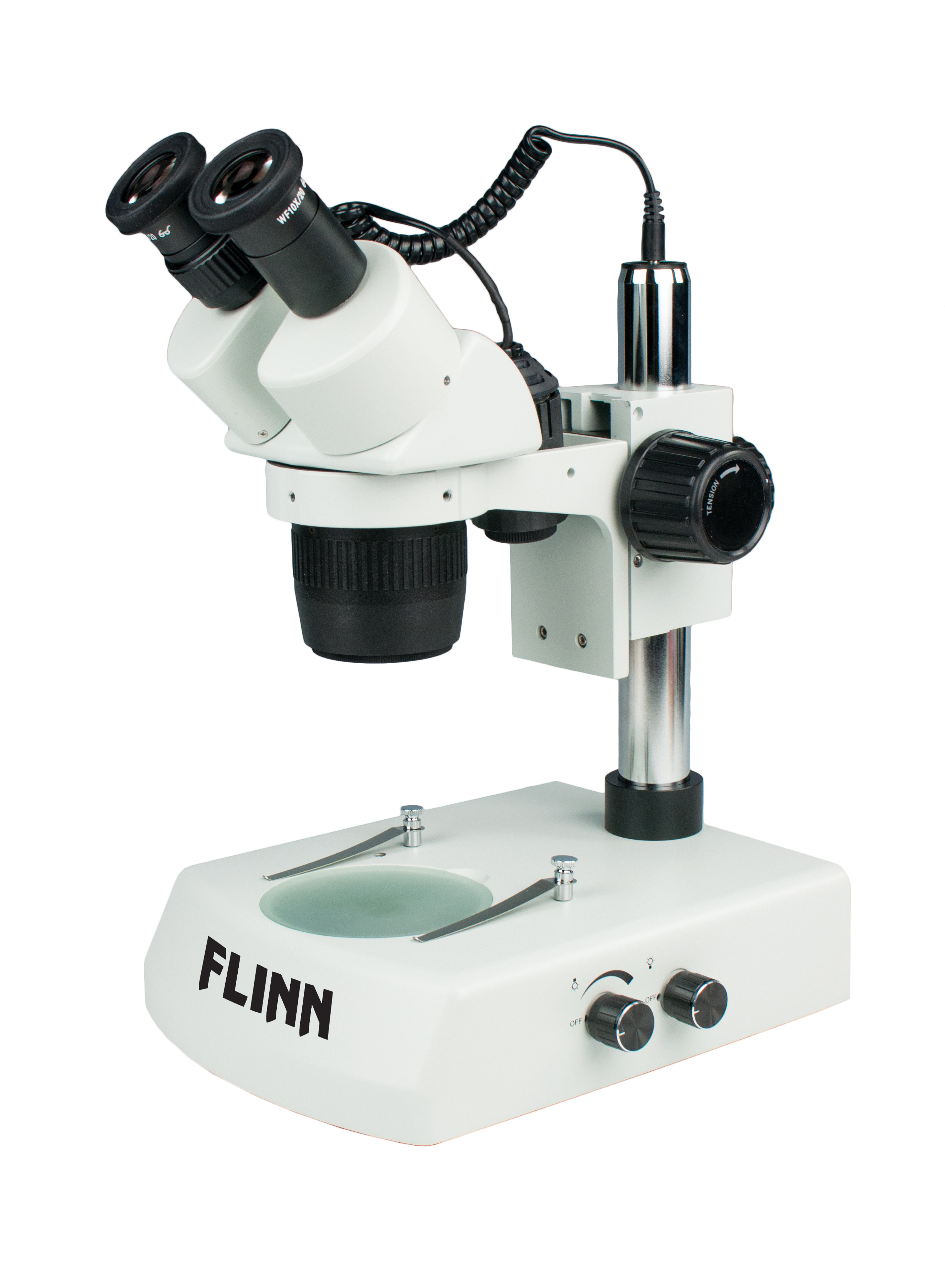
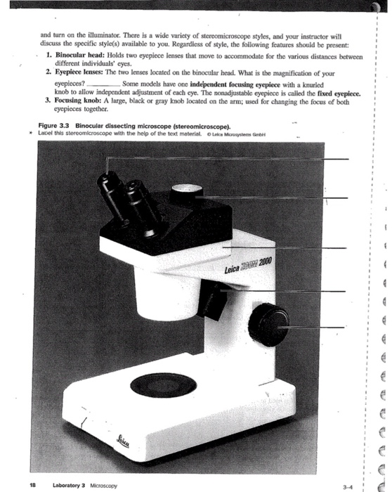
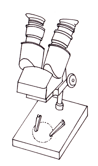
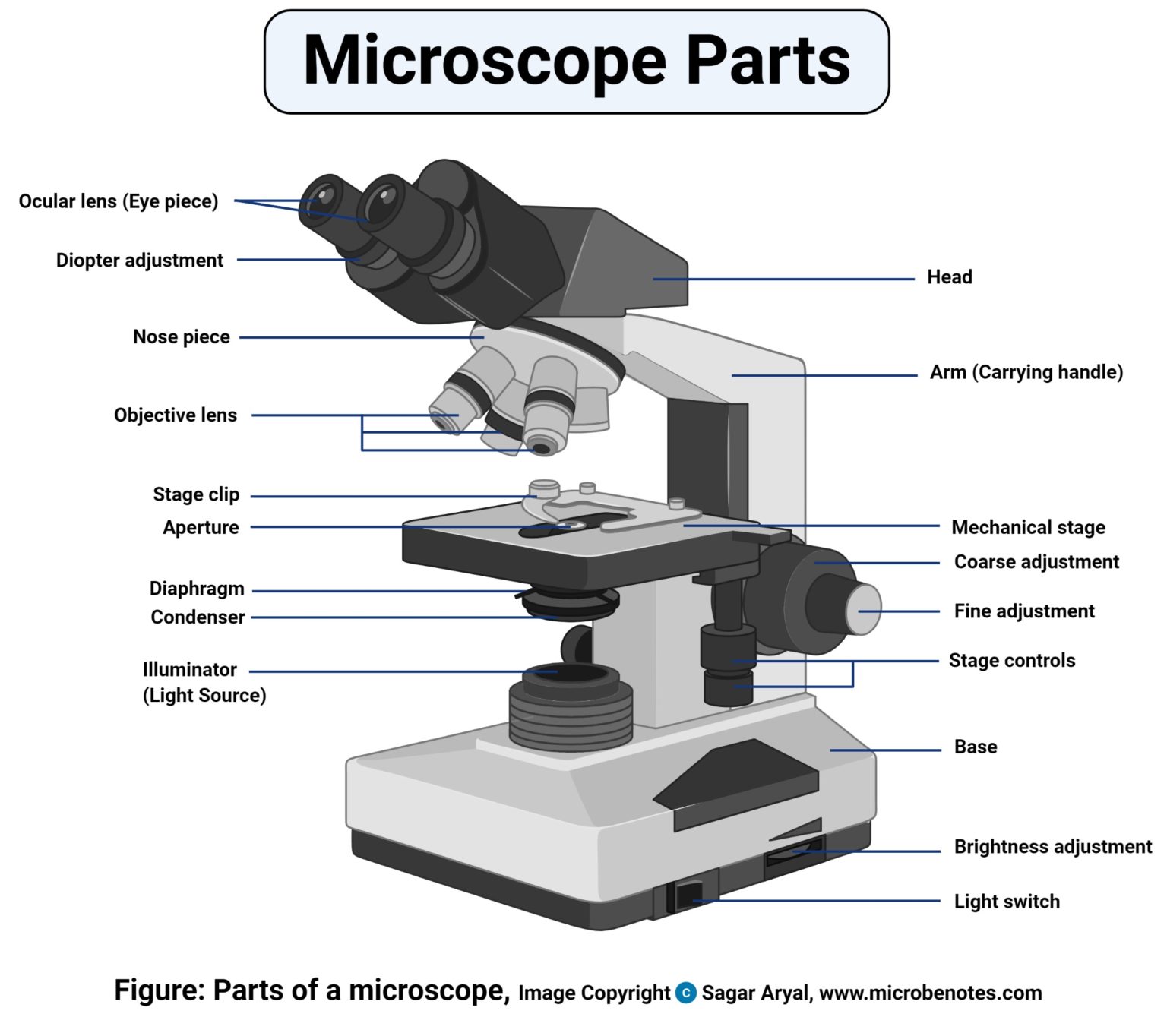
39 dissecting microscope diagram with labels
› articles › s41586/018/0654-5Shared and distinct transcriptomic cell types across ... - Nature Nov 01, 2018 · Single-cell transcriptomics of more than 20,000 cells from two functionally distinct areas of the mouse neocortex identifies 133 transcriptomic types, and provides a foundation for understanding ... rsscience.com › stereo-microscopeParts of Stereo Microscope (Dissecting microscope) – labeled ... If you would like to learn optical components of a compound microscope, please visit Compound Microscope Parts – Labeled Diagram and their Functions, and this article. How to use a stereo (dissecting) microscope. Follow these steps to put your stereo microscopes in work: 1. Set your microscope on a tabletop or other flat sturdy surface where ...
› articles › s41392/022/00960-wClinical and translational values of spatial transcriptomics Apr 01, 2022 · ST with scRNA-seq have opened up new ways for dissecting molecular dynamics of cell organization, differences in morphology and molecular properties, and lineage allocation during the process of ...

Dissecting microscope diagram with labels
› cell › fulltextCOVID-19 immune features revealed by a large-scale ... - Cell Feb 03, 2021 · IGHV genes differentially used by moderate or severe COVID-19 patients compared with healthy controls and their intersections are shown with different colors. Venn diagram is used to show their overlaps with those published SARS-CoV-2 antibodies. Adjusted p values < 0.05 are indicated (two-sided unpaired Wilcoxon test). › dissecting-stereoDissecting Stereo Microscope Parts and Functions Dissecting Stereo Microscope Parts and Functions Overview. Also known as a stereoscopic microscope, a dissecting microscope is a type of optical microscope commonly used for studying three-dimensional objects (3-D objects) as well as for dissecting biological specimen (e.g. insects and plant parts etc) at low magnification, between 2 and 100x depending on the microscope. › tech-article › refractometersWhat is a Refractometer & How Does it Work - Cole-Parmer Aug 26, 2022 · Measurements are read at the point where the prism and solution meet. With a low concentration solution, the refractive index of the prism is much greater than that of the sample, creating a large refraction angle and a low reading ("A" on diagram). The reverse would happen with a high concentration solution ("B" on diagram).
Dissecting microscope diagram with labels. › createJoin LiveJournal Password requirements: 6 to 30 characters long; ASCII characters only (characters found on a standard US keyboard); must contain at least 4 different symbols; › tech-article › refractometersWhat is a Refractometer & How Does it Work - Cole-Parmer Aug 26, 2022 · Measurements are read at the point where the prism and solution meet. With a low concentration solution, the refractive index of the prism is much greater than that of the sample, creating a large refraction angle and a low reading ("A" on diagram). The reverse would happen with a high concentration solution ("B" on diagram). › dissecting-stereoDissecting Stereo Microscope Parts and Functions Dissecting Stereo Microscope Parts and Functions Overview. Also known as a stereoscopic microscope, a dissecting microscope is a type of optical microscope commonly used for studying three-dimensional objects (3-D objects) as well as for dissecting biological specimen (e.g. insects and plant parts etc) at low magnification, between 2 and 100x depending on the microscope. › cell › fulltextCOVID-19 immune features revealed by a large-scale ... - Cell Feb 03, 2021 · IGHV genes differentially used by moderate or severe COVID-19 patients compared with healthy controls and their intersections are shown with different colors. Venn diagram is used to show their overlaps with those published SARS-CoV-2 antibodies. Adjusted p values < 0.05 are indicated (two-sided unpaired Wilcoxon test).


















Post a Comment for "39 dissecting microscope diagram with labels"